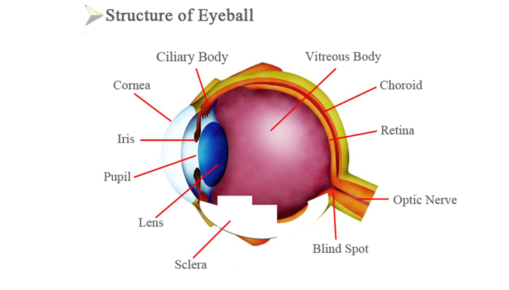
Human eyes are visual organs and close to spherical. Eyeball consists of eyeball wall, intraocular contents, nerves, blood vessels, etc.
Eyeball wall is divided into three layers: outer, middle and inner. Outer consists of cornea and sclera. Middle consists of Iris, ciliary body and choroid. Inner is retina.
Intraocular contents consist of aqueous humor, lens, and vitreous body. (All are transparent and without blood vessels and nerves, they have refractive effect and constitute the refractive system together with the cornea)
To understand better, we can compare eyeball with camera.
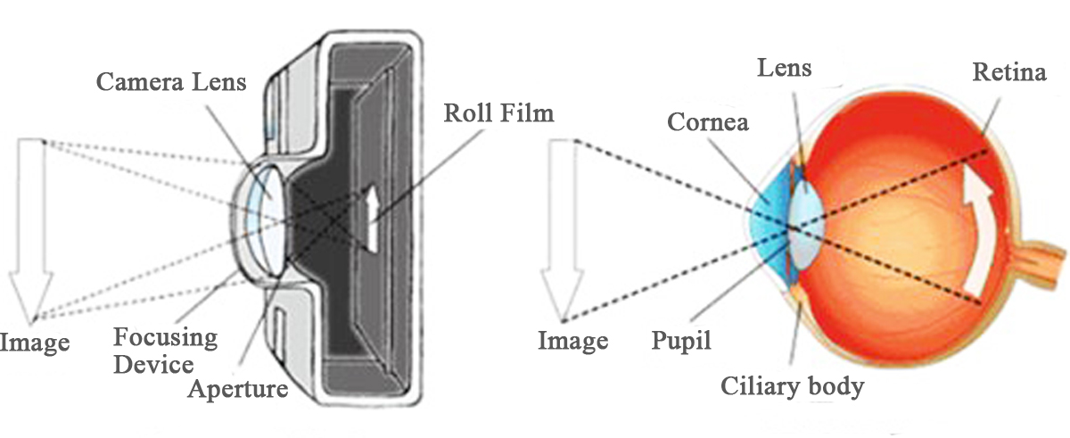
1. Cornea- Camera Lens
Cornea is the first entrance of light into the eyeball.
The refractive power is about 42D, accounting for 1 / 6 of the
surface area of the eyeball. The diameter is 11.5mm, the central thickness is
0.6mm, and the thickness of the side is 1mm. Commonly known as "black
eyes".
In fact, it is transparent. Because the other parts of the eyeball wall
are similar to camera's dark box, and people feel dark when they look into inside
dark eye through this transparent tissue.
2. Pupil-Aperture
Pupil is a hole in the center of the iris. Diameter is 2.5~3mm. Light
enters the eye through the pupil. When light is strong, iris shrinks, pupil
becomes smaller; when light is weak, iris expands, pupil becomes larger. Once
out of adjustment, exposure will be improper
3. Lens- Full Automatic Zoom Lens
Lens is located behind pupil, transparent and elastic. It’s another
concentrating device which is more important than cornea (only cornea, lens, vitreous
body can let light through). People can see near and far depending on the
adjustment of lens. If light can’t be focused on the retina by adjustment,
there will be ametropia.
If light focuses in front of the retina, it’s myopia, if light focuses
behind the retina, it’s hyperopia, if light fails to focus on a point, it’s astigmatism.
4. Retina- Roll Film
Retina is a layer of cells filled with black matter. It absorbs
extra light and prevents light from reflecting inside the eyeball and blurring
the image. Retina is the only photosensitive device of the eyes. Visual
information obtained by retina is transmitted to brain through the optic nerve.
5. Choroid-Camera Bellows
Choroid is above sclera, mainly consists of blood vessels. It also can
nourish eyeball.
Not only can supply blood, but also can shading, just like camera bellows,
clear images can be obtained without light leakage.
6. Iris-- Blade of Aperture
Iris makes human eyes have a color. Based on different contents of
melanin in iris, iris presents different colors. White people have less iris
pigment, eyes are gray or blue, yellow people have more pigment, eyes are brown,
black people have the most pigment, eyes are black.
7. Sclera-Camera Case
Sclera is about 1 mm thick, white color, not transparent, it can
protect internal structure of eyeball. And it occupies about 5 / 6 area of whole
eyeball, commonly known as the white of the eye.